
Mouse Secondary Antibodies


Biotium Spectrin beta III (SPTBN2) (SPTBN2/1247), CF568 conjugate, 0.1mg/mL
Spectrin is an actin binding protein that is a major component of the plasma membrane skeleton. Spectrins function as membrane organizers and stabilizers by forming dimers, tetramers and higher polymers. Vertebrate spectrins have two alpha-subunits (alpha-I/alpha-II), four beta-subunits (beta-I-beta-IV) and a beta-H subunit creating diversity and specialization of function. Spectrin alpha and spectrin beta are present in erythrocytes, whereas spectrin alpha II (also designated fodrin alpha) and spectrin beta I (also designated fodrin beta) are present in other somatic cells. The spectrin tetramers in erythrocytes act as barriers to lateral diffusion, but spectrin dimers seem to lack this function. Spectrin beta III is highly homologous to both spectrin beta I and spectrin beta II. Spectrin beta III is highly expressed in brain, kidney, pancreas and liver, and at lower levels in lung and placenta. Spectrin beta 3 is primarily expressed in nervous tissues with highest expressio

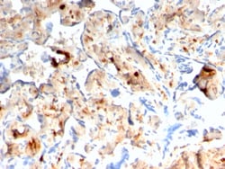
Biotium S100A4 / Metastasin / Calvasculin (Marker of Tumor Metastasis)(rS100A4/1481), CF740 conjugate, 0.1mg/mL
S100A4 belongs to the S100 super-family of proteins containing 2 EF-hand calcium-binding domains. S100 genes include at least 25 members, including S100A1-S100A18, trichohyalin, filaggrin, repetin, S100P, and S100Z. S100A4 exerts its function via direct interaction with a number of proteins including P53, P63, non-muscle myosin IIA, 64 integrin, and liprin b1. S100A4 is overexpressed in highly metastatic cancers, which makes it useful as a marker of tumor progression. Primary antibodies are available purified, or with a selection of fluorescent CF Dyes and other labels. CF Dyes offer exceptional brightness and photostability. Note: Conjugates of blue fluorescent dyes like CF405S and CF405M are not recommended for detecting low abundance targets, because blue dyes have lower fluorescence and can give higher non-specific background than other dye colors.
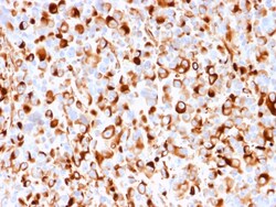
Biotium NGF-Receptor (p75) / CD271 (Soft Tissue Tumor Marker)(rNGFR/1965), CF488A conjugate, 0.1mg/mL
Recognizes a glycoprotein of 75 kDa, identified as low affinity Nerve Growth Factor (NGF) Receptor (p75NGFR) or Neurotrophin Receptor (p75NTR). NGFR is expressed in various neural crest cells and their tumors such as melanocytes, melanomas, neuroblastomas, pheochromocytomas and neurofibromas. Reportedly, anti-NGFR is a reliable marker for desmoplastic and neurotropic melanomas. NGFR is expressed in mature non-neural cells such as perivascular cells, dental pulp cells, lymphoidal follicular dendritic cells, basal epithelium of oral mucosa and hair follicles, prostate basal cells, and myoepithelial cells. Anti-NGFR stains the myoepithelial cells of breast ducts and intra-lobular fibroblasts of breast ducts. Primary antibodies are available purified, or with a selection of fluorescent CF Dyes and other labels. CF Dyes offer exceptional brightness and photostability. Note: Conjugates of blue fluorescent dyes like CF405S and CF405M are not recommended for detecting low abunda
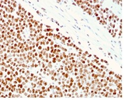
Biotium MSH6 (DNA Mismatch Repair Protein)(MSH6/3086), CF740 conjugate, 0.1mg/mL
The finding that mutations in DNA mismatch repair genes are associated with hereditary nonpolyposis colorectal cancer (HNPCC) has resulted in considerable interest in the understanding of the mechanism of DNA mismatch repair. Initially, inherited mutations in the MSH2 and MLH1 homologs of the bacterial DNA mismatch repair genes mutS and mutL were demonstrated at high frequency in HNPCC and were shown to be associated with microsatellite instability. A member of the mismatch repair family, GTBP (also designated MSH6), is an MSH2-related protein that binds to DNA containing G/T mismatches. Findings suggest that the mismatch-binding factor in human cells is composed of a heterodimer of GTBP and MSH2. Primary antibodies are available purified, or with a selection of fluorescent CF Dyes and other labels. CF Dyes offer exceptional brightness and photostability. Note: Conjugates of blue fluorescent dyes like CF405S and CF405M are not recommended for detecting low abundance targ
Biotium Spectrin Alpha 1 (Erythrocyte Marker) (SPTA1/2939R), CF488A conjugate, 0.1mg/mL
Spectrin is an actin crosslinking and molecular scaffold protein that links the plasma membrane to the actin cytoskeleton, and functions in the determination of cell shape, arrangement of transmembrane proteins, and organization of organelles. Spectrins function as membrane organizers and stabilizers, composed of non-homologous alpha and beta chains, which aggregate side-to-side in an anti-parallel fashion to form dimers, tetramers and higher polymers. Spectrin alpha I and spectrin beta I are present in erythrocytes, whereas spectrin alpha II (also designated fodrin alpha) and spectrin beta II (also designated fodrin beta) are present in other somatic cells. The spectrin tetramers in erythrocytes act as barriers to lateral diffusion, but spectrin dimers seem to lack this function.Primary antibodies are available purified, or with a selection of fluorescent CF Dyes and other labels. CF Dyes offer exceptional brightness and photostability. Note: Conjugates of blue fluorescent dye
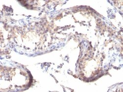
Biotium TGFalpha(TGFA/1119), CF568 conjugate, 0.1mg/mL
This antibody reacts with the C-terminus of TGF alpha and shows no cross-reaction with EGF and the neuropeptide synenkephalin. TGF-beta (aa50) is a growth factor with 33% homology to EGF, binds to EGFR, activates tyrosine phosphorylation of the receptor, and stimulates cell proliferation. It plays a role in tumor initiation by inducing the reversible transformed phenotype.Primary antibodies are available purified, or with a selection of fluorescent CF Dyes and other labels. CF Dyes offer exceptional brightness and photostability. Note: Conjugates of blue fluorescent dyes like CF405S and CF405M are not recommended for detecting low abundance targets, because blue dyes have lower fluorescence and can give higher non-specific background than other dye colors.
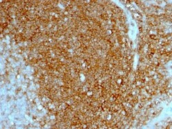
Biotium CD44 Standard(HCAM/1097), CF568 conjugate, 0.1mg/mL
Recognizes a cell surface glycoprotein of 80-95 kDa (CD44) on lymphocytes, monocytes, and granulocytes. The CD44 family of glycoproteins exists in a number of variant isoforms, the most common being the standard 85-95 kDa or hematopoietic variant (CD44s). Higher molecular weight isoforms are described in epithelial cells (CD44v), which are believed to function in intercellular adhesion and stromal binding. CD44 immunostaining is commonly used for the discrimination of urothelial transitional cell carcinoma in-situ from non-neoplastic changes in the urothelium.Primary antibodies are available purified, or with a selection of fluorescent CF Dyes and other labels. CF Dyes offer exceptional brightness and photostability. Note: Conjugates of blue fluorescent dyes like CF405S and CF405M are not recommended for detecting low abundance targets, because blue dyes have lower fluorescence and can give higher non-specific background than other dye colors.
Biotium von Willebrand Factor / Factor VIII Related-Ag (Endothelial Marker)(IIIE2.34), CF740 conjugate, 0.1mg/mL
von Willebrand Factor (vWF) is a multimeric glycoprotein that is found in endothelial cells, plasma and platelets. It acts as a carrier protein for Factor VIII and promotes platelet adhesion and aggregation. vWF undergoes a variety of posttranslational modifications that influence the affinity and availability for Factor VIII, including cleavage of the propeptide and formation of N-terminal disulfide bonds. This antibody helps to establish the endothelial nature of some lesions of disputed histogenesis, e.g. Kaposi's sarcoma and cardiac myxoma. It is widely used for differentiating vascular lesions from those of other tissue differentiation within a panel of other vascular markers although not all tumors of endothelial differentiation contain this antigen.
Biotium CD7(124.1D1), CF488A conjugate, 0.1mg/mL
Recognizes a protein of 40 kDa, identified as CD7 (Workshop IV; Code T155). CD7 is a member of the immunoglobulin gene superfamily. Its N-terminal amino acids 1-107 are highly homologous to Ig kappa-L chains whereas the carboxyl-terminal region of the extracellular domain is proline-rich and has been postulated to form a stalk from which the Ig domain projects. CD7 is expressed on the majority of immature and mature T-lymphocytes, and T cell leukemia. It is also found on natural killer cells, a small subpopulation of normal B cells and on malignant B cells. Cross-linking surface CD7 positively modulates T cell and NK cell activity as measured by calcium fluxes, expression of adhesion molecules, cytokine secretion and proliferation. CD7 associates directly with phosphoinositol 3'-kinase. CD7 ligation induces production of D-3 phosphoinositides and tyrosine phosphorylation.Primary antibodies are available purified, or with a selection of fluorescent CF Dyes and other labels. C
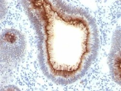
Biotium CD66 (CEA)(C66/1009), CF740 conjugate, 0.1mg/mL
This antibody recognizes proteins of 80-200 kDa, identified as different members of CEA family. CEA is synthesized during development in the fetal gut and is re-expressed in increased amounts in intestinal carcinomas and several other tumors. This MAb does not react with nonspecific cross-reacting antigen (NCA) and with human polymorphonuclear leucocytes. It shows no reaction with a variety of normal tissues and is suitable for staining of formalin/paraffin tissues. CEA is not found in benign glands, stroma, or malignant prostatic cells. Antibody to CEA is useful in detecting early foci of gastric carcinoma and in distinguishing pulmonary adenocarcinomas (60-70% are CEA positive) from pleural mesotheliomas (rarely or weakly CEA positive). Anti-CEA positivity is seen in adenocarcinomas from the lung, colon, stomach, esophagus, pancreas, gallbadder, urachus, salivary gland, ovary, and endocervix.Primary antibodies are available purified, or with a selection of fluorescent CF
Biotium MUC5AC (Mucin 5AC / Gastric Mucin)(9-13M1), 0.2mg/mL
This MAb recognizes the peptide core of gastric mucin M1 (recently identified as Mucin 5AC). Its epitope is located in the N-terminal cysteine rich part of the peptide core of MUC5AC, which is heavily glycosylated. Its epitope is destroyed by beta-mercaptoethanol but not by periodate treatment. MAb 2-11M1 reacts with the protein backbone exclusively; it only reacts with fully deglycosylated MUC5AC. Therefore, the material under test should also be fully deglycosylated. This can be achieved with standard periodate oxidation method. The success of the deglycosylation can be checked with routine PAS (Periodic Acid Shiff) staining. After deglycosylation, the preparation should no longer be stainable with PAS reagent. Only then 2-11M1 will react should MUC5AC be present. This mucin is present in primary ovarian mucinous cancer but usually absent in colorectal adenocarcinoma, thus showing an expression pattern opposite to MUC2. Together with a panel of antibodies, Anti-MUC5AC may b
Biotium Spectrin beta III (SPTBN2) (SPTBN2/1247), Biotin conjugate, 0.1mg/mL
Spectrin is an actin binding protein that is a major component of the plasma membrane skeleton. Spectrins function as membrane organizers and stabilizers by forming dimers, tetramers and higher polymers. Vertebrate spectrins have two alpha-subunits (alpha-I/alpha-II), four beta-subunits (beta-I-beta-IV) and a beta-H subunit creating diversity and specialization of function. Spectrin alpha and spectrin beta are present in erythrocytes, whereas spectrin alpha II (also designated fodrin alpha) and spectrin beta I (also designated fodrin beta) are present in other somatic cells. The spectrin tetramers in erythrocytes act as barriers to lateral diffusion, but spectrin dimers seem to lack this function. Spectrin beta III is highly homologous to both spectrin beta I and spectrin beta II. Spectrin beta III is highly expressed in brain, kidney, pancreas and liver, and at lower levels in lung and placenta. Spectrin beta 3 is primarily expressed in nervous tissues with highest expressio
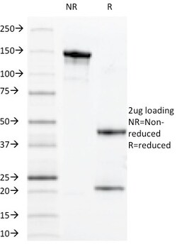
Biotium MUC1 / CA15-3 / EMA / CD227 (Epithelial Marker)(VU-11D1), CF740 conjugate, 0.1mg/mL
MAb VU-11D1 reacts with MUC1, a large transmembrane glycoprotein expressed on the ductal surface of normal glandular epithelia. The dominant epitope of MAb VU-11D1 is the 7-mer TSAPDTR of the MUC1 tandem repeat as established with 'epitope fingerprinting'. The'extra cellular domain of MUC1 largely consists of a highly conserved, O-glycosylated 20 amino acids tandem repeat which can occur 30-100 times per molecule depending on the length of the allele involved. In the vast majority of human carcinomas this protein is up regulated and poorly glycosylated and appears on the cell surface in a non-polarized fashion. Primary antibodies are available purified, or with a selection of fluorescent CF Dyes and other labels. CF Dyes offer exceptional brightness and photostability. Note: Conjugates of blue fluorescent dyes like CF405S and CF405M are not recommended for detecting low abundance targets, because blue dyes have lower fluorescence and can give higher non-specific backgrou
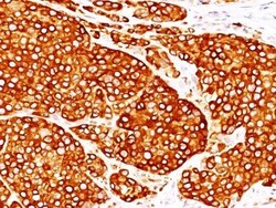
Biotium Tyrosinase(T311 + OCA1/812), CF740 conjugate, 0.1mg/mL
Recognizes a cluster of proteins between 70-80 kDa, identified as tyrosinase. Occasionally a minor band at 55 kDa is also detected. This MAb shows no cross-reaction with MAGE-1 and tyrosinase-related protein 1, TRP-1/gp75. Tyrosinase is a copper-containing metalloglycoprotein that catalyzes several steps in the melanin pigment biosynthetic pathway; the hydroxylation of tyrosine to L-3,4-dihydroxy-phenylalanine (dopa), and the subsequent oxidation of dopa to dopaquinone. Mutations of the tyrosinase gene occur in various forms of albinism. Tyrosinase is one of the targets for cytotoxic T-cell recognition in melanoma patients. Staining of melanomas with this MAb shows tyrosinase in melanotic as well as amelanotic variants. This MAb is a useful marker for melanocytes and melanomas.Primary antibodies are available purified, or with a selection of fluorescent CF Dyes and other labels. CF Dyes offer exceptional brightness and photostability. Note: Conjugates of blue fluorescent d
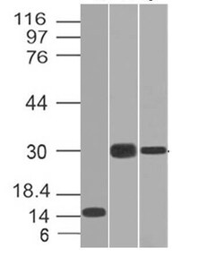
Biotium CELA3B(CELA3B/1218), Biotin conjugate, 0.1mg/mL
This MAb recognizes a protein of ~17 kDa, identified as CELA3B (Chymotrypsin like elastase family member 3B). Elastases form a subfamily of serine proteases that hydrolyze many proteins in addition to elastin. Humans have six elastase genes which encode the structurally similar proteins elastase 1, 2, 2A, 2B, 3A, and 3B. Unlike other elastases, elastase 3B has little elastolytic activity. Like most of the human elastases, elastase 3B is secreted from the pancreas as a zymogen and, like other serine proteases such as trypsin, chymotrypsin and kallikrein; it has a digestive function in the intestine. Elastase 3B preferentially cleaves proteins after alanine residues. Elastase 3B may also function in the intestinal transport and metabolism of cholesterol. Both elastase 3A and elastase 3B have been referred to as protease E and as elastase 1, and excretion of this protein in fecal material is frequently used as a measure of pancreatic function in clinical assays.Primary antibodie